Back جهاز بطيني Arabic Мозъчни вентрикули Bulgarian Kambr (empenn) Breton Sistema ventricular Catalan Mozková komora Czech Ventrikelsystem Danish Hirnventrikel German Ventrículo cerebral Spanish Ajuvatsakesed Estonian دستگاه بطنی Persian
| Ventricular system | |
|---|---|
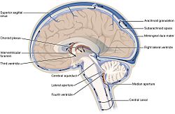
 The ventricular system accounts for the production and circulation of cerebrospinal fluid. | |
 Rotating 3D rendering of the four ventricles and connections. From top to bottom: Blue - lateral ventricles Cyan - interventricular foramina (Monro) Yellow - third ventricle Red - cerebral aqueduct (Sylvius) Purple - fourth ventricle Green - continuous with the central canal (Parts between median aperture and subarachnoid space are not shown) | |
| Details | |
| Identifiers | |
| Latin | ventriculi cerebri |
| MeSH | D002552 |
| NeuroNames | 2497 |
| FMA | 242787 |
| Anatomical terms of neuroanatomy | |
In neuroanatomy, the ventricular system is a set of four interconnected cavities known as cerebral ventricles in the brain.[1][2] Within each ventricle is a region of choroid plexus which produces the circulating cerebrospinal fluid (CSF). The ventricular system is continuous with the central canal of the spinal cord from the fourth ventricle,[3] allowing for the flow of CSF to circulate.[3][4]
All of the ventricular system and the central canal of the spinal cord are lined with ependyma, a specialised form of epithelium connected by tight junctions that make up the blood–cerebrospinal fluid barrier.[2]
- ^ Grow, W.A. (2018). "Development of the Nervous System". Fundamental Neuroscience for Basic and Clinical Applications. Elsevier. pp. 72–90.e1. doi:10.1016/b978-0-323-39632-5.00005-0. ISBN 978-0-323-39632-5.
The ventricular system is an elaboration of the lumen of cephalic portions of the neural tube, and its development parallels that of the brain.
- ^ a b Shoykhet, Mish; Clark, Robert S.B. (2011). "Structure, Function, and Development of the Nervous System". Pediatric Critical Care. Elsevier. pp. 783–804. doi:10.1016/b978-0-323-07307-3.10057-6. ISBN 978-0-323-07307-3.
The ventricles contain the choroid plexus, which produces CSF, and serve as conduits for CSF flow in the CNS. Ventricular walls are lined with ependymal cells, which are connected by tight junctions and constitute a CSF-brain barrier.
- ^ a b Shoykhet, Mish; Clark, Robert S.B. (2011). "Structure, Function, and Development of the Nervous System". Pediatric Critical Care. Elsevier. pp. 783–804. doi:10.1016/b978-0-323-07307-3.10057-6. ISBN 978-0-323-07307-3.
The ventricular system arises from the hollow space within the developing neural tube and gives rise to cisterns within the CNS, from the brain to the spinal cord.
- ^ Vernau, William; Vernau, Karen A.; Sue Bailey, Cleta (2008). "Cerebrospinal Fluid". Clinical Biochemistry of Domestic Animals. Elsevier. pp. 769–819. doi:10.1016/b978-0-12-370491-7.00026-x. ISBN 978-0-12-370491-7. S2CID 71013935.
Cerebrospinal fluid flows in bulk from sites of production to sites of absorption. Fluid formed in the lateral ventricles flows through the paired interventricular foramina (foramen of Monro) into the third ventricle, then through the mesencephalic aqueduct (aqueduct of Sylvius) into the fourth ventricle. The majority of CSF exits from the fourth ventricle into the subarachnoid space; a small amount may enter the central canal of the spinal cord.
© MMXXIII Rich X Search. We shall prevail. All rights reserved. Rich X Search